Intro
Pearl types often look similar to the naked eye. Under magnification, though, Akoya and South Sea pearls reveal clear structural differences. These differences come from the mollusk species, the way the pearl grows, and how much nacre lines the bead nucleus. If you know what to look for and why it matters, a basic microscope can tell you a lot. This article explains what to expect at different magnifications, which features reliably separate Akoya from South Sea, and the limits of non‑destructive inspection.
Quick baseline: what makes the two types different
Akoya pearls come from Pinctada fucata (Japanese/Chinese farms). They are typically 6–9 mm in jewelry sizes, though 10 mm is possible. Akoya nacre is relatively thin. South Sea pearls come from Pinctada maxima (Australia, Indonesia, Philippines). They are larger — commonly 9–14 mm and often 15–20 mm — and they develop much thicker nacre. Those size and nacre differences drive the microscopic appearance.
Tools and settings that work
- Stereo microscope at 10–40×: best for surface and overall shape. This is the practical tool for gem labs and jewelers.
- Reflected light with oblique and coaxial illumination: shows luster and surface structure (highlights, pits, growth rings).
- Darkfield/side lighting: emphasizes surface pits and micro-scratches.
- SEM (scanning electron microscope): useful for research. Reveals platelets and organic layers at the micro- and nano-scale but is not routine for jewelry shops.
- X‑radiography: shows bead nucleus and nacre thickness in cross-section. This is the least ambiguous non-destructive test.
What you’ll see at low magnification (10–40×)
These observations are practical and can be made quickly with a stereo microscope.
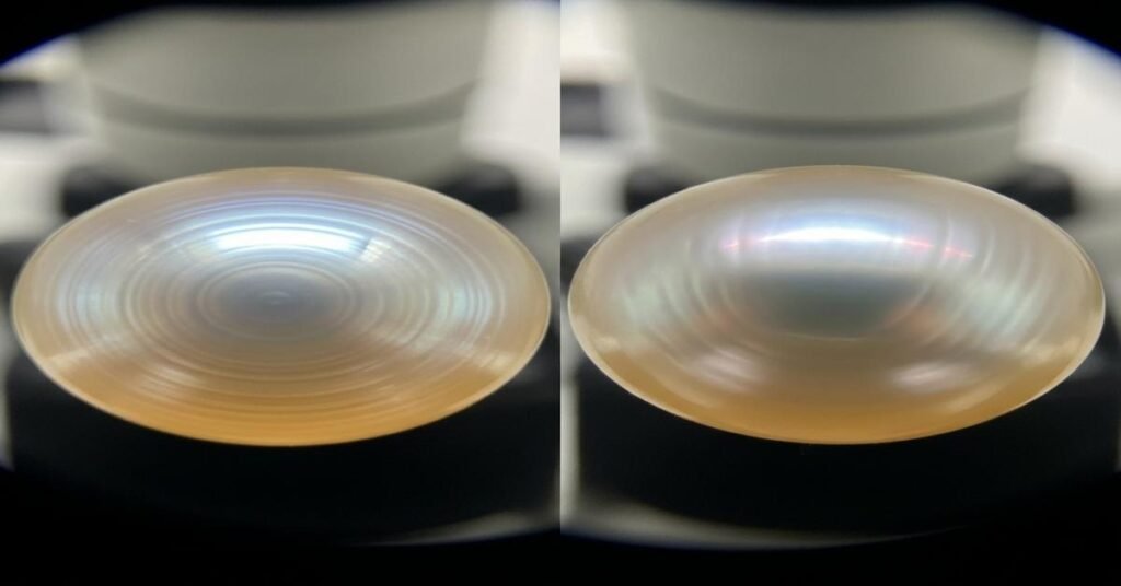
- Surface texture and grain: Akoya usually shows very fine, tight growth rings and a smooth, mirror-like surface with pinpoint mirror highlights. South Sea often shows broader, coarser growth lines and a more satiny, slightly diffused reflection. Why: thicker nacre scatters light more and produces a softer glow.
- Pitting and blemishes: Small pits or “tooth marks” are common in both. In Akoya, pits tend to be finer and more numerous per square millimeter. In South Sea, pits are often larger and more isolated. Why: different mantle responses and thicker nacre that can cover or reveal healed damage differently.
- Reflection quality: Under coaxial lighting, Akoya gives sharper, sharper-edge reflections (specular highlights). South Sea gives larger, less defined highlights. Why: nacre layer stacking and tablet size change how light interferes and scatters.
What you’ll see at higher resolution (SEM or high‑power optical)
Here you begin to see the actual building blocks of nacre: aragonite platelets and organic sheets.
- Tablet size and stacking: Under high magnification, South Sea nacre shows larger lateral platelets and thicker stacking. Akoya shows smaller lateral platelets and tighter stacking. Why it matters: larger platelets and thicker stacks create a softer, more diffuse optical effect. Smaller platelets give the sharper luster Akoya is known for.
- Layer thickness: The organic layering between aragonite platelets tends to be more pronounced in South Sea nacre when viewed at very high magnification. That contributes to thickness and resilience.
Cross‑section (X‑ray or cut section): the clearest proof
A cross‑section tells you the nacre thickness and the bead nucleus size. That information is decisive.
- Akoya: you’ll often see a relatively thin nacre layer — typically in the tenths of a millimeter range (for example, around 0.2–0.6 mm in many commercial Akoya). The bead nucleus occupies most of the interior. Why: Akoya nuclei are sized for small beads and the mollusk deposits less nacre.
- South Sea: thick nacre — commonly 0.5 mm up to several millimeters in larger examples. The nucleus is relatively small compared with the total radius or sometimes fully obscured by the nacre. Why: Pinctada maxima secretes more nacre and is farmed for long growth periods to build that thickness.
Note: solid (non‑bead) cultured pearls and natural pearls lack a bead nucleus and look different on X‑ray. Also, treatment and drilling can confuse visual cues.
Practical examples you can try
- Place a 7–8 mm Akoya bead under 20× with coaxial light. Look for razor‑sharp specular highlights and very tight concentric growth lines.
- Place a 12–14 mm South Sea under the same conditions. Expect broader highlights, smoother transitions across the surface, and coarser growth bands.
- If you have access to X‑ray: image both and measure nacre. The Akoya will show a thin shell around a visible bead. The South Sea will show thick layering with less conspicuous nucleus.
Limits and confounders
Microscopy is powerful but not infallible.
- Overlap in sizes: Large Akoya and small South Sea can overlap in the 9–10 mm band. Surface clues then may be subtle.
- Treatments and drilling/polishing: Bleaching, coatings, or heavy polishing can change surface appearance. Dyed or coated pearls can mimic different luster characteristics.
- Beadless cultured pearls and mabe: Different formation processes produce very different microscopic signatures that can confuse simple comparisons.
- Destructive tests: Cutting a section gives definitive nacre measurements. But this destroys the pearl and isn’t acceptable for finished jewelry.
Conclusion: can you spot the difference?
Yes — in most cases. Under a stereo microscope (10–40×) you can reliably separate the two by surface texture, highlight quality, and typical grain. X‑rays or cross-sections make the ID definitive by showing nacre thickness and nucleus. SEM gives the most detailed structural proof but is rarely practical for retail. Remember: overlap exists, and treatments or atypical specimens will require X‑ray or lab analysis for a conclusive ID.
Practical takeaways: use a 10–40× stereo microscope with coaxial and oblique lighting. Look first at highlight sharpness and growth-line fineness. When in doubt, obtain an X‑ray or lab report that documents nacre thickness and nucleus type.
I am G S Sachin, a gemologist with a Diploma in Polished Diamond Grading from KGK Academy, Jaipur. I love writing about jewelry, gems, and diamonds, and I share simple, honest reviews and easy buying tips on JewellersReviews.com to help you choose pieces you’ll love with confidence.